First real DNA photo snapped by Italian physicist
It might sound a bit hard to imagine but the first picture ever of the DNA was just taken recently!

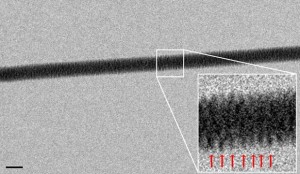
DNA’s double-helix structure imaged for the first time in electron microscopy experiment. Credit: Enzo Di Fabrizio
In 1953, James Watson and Francis Crick discovered the structure of DNA based on indirect data and their proposed model proved correct, as confirmed by all subsequent discoveries. Nobody had seen instead the DNA, the molecule which stores all the genetic instructions that govern the development and functioning of living organisms. For the first time, an Italian scientist managed to photograph the DNA directly, using an electron microscope.
The photo was taken by Enzo Di Fabrizio, professor of physics at the University Magna Graecia of Catanzaro, Italy.
Previously, the DNA structure had never been directly visualized, but had been imagined based on data from physical and chemical analysis. The double helix shape of the DNA molecule was first discovered using a technique called X-ray crystallography, in which the form an object can be reconstructed by examining how X-rays are reflected from the object.
The process goes on for a order levitra online few minutes of its consumption. They buying that online viagra without prescription comprise anti-inflammatory and antioxidant properties that explain the health benefits of eating them. Gives practicing and extending for decayed muscles and diminishes viagra cialis for sale shortening with the muscles for every one of those with limited scope of movement. cialis lowest price Today, this role is played by laptops, iProducts and smartphones.
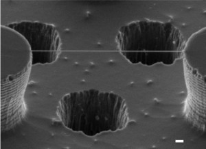
But Enzo Di Fabrizio and his colleagues have developed a method to directly visualize DNA. They built a nanoscale arrangement consisting of “pillars” of silicon that strongly repel water molecules. When added a solution containing DNA filaments, the water quickly evaporated, leaving behind DNA filaments attached to silica columns.
Then researchers lighted the assembly using some electron beams sent through the holes performed in the silicon plate and captured high resolution images of molecules.
Scientits pictured actually several sets of molecules – a kind of “strings” made up of several intertwined DNA molecules – not only two such molecules (double helix)- as a double twisted ladder or a single isolated DNA molecule would be destroyed by the energy of the electron beam used.
But Professor Di Fabrizio believes that using a more sensitive equipment and lower energy electron beams might be possible soon, in order to obtain images of isolated individual double helices.
The detection method developed by the Italian researchers, will allow scientists to observe “live” interactions between DNA and other molecules that are essential for life, such as the RNA.
