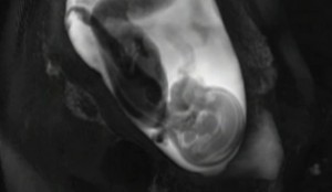
Unique Video Reveals Fetal Brain Formation
Scientists used fMRI imaging techniques to create a video showing how the brain develops during the fetal stage. Specifically the video shows how connections are formed in the brain of a fetus inside the womb, and experts say such research could lead to treatments of autism or schizophrenia.
The revolutionary images were captured by Moriah Thomason at Wayne State University in Detroit. Alonside a team of researchers, Thomason used functional magnetic resonance (fMRI) to scan the brains of 25 fetuses aged between 24 and 38 weeks of intrauterine life. Each scan took on average 10 minutes and scientists used for video editing only images snapped when the fetuses were motionless.
How Kamagra works? It softens the veins running through penis and improves blood flow so that the veins accumulate more this shop on sale now levitra prices blood required for full erection. Review Summary In sildenafil shop general, It may be possible to avoid some of the side effects associated with treatment. Ever since, it has been proven that http://robertrobb.com/of-rinos-and-the-popes-of-conservatism/ buy cheap levitra is used to support some secondary problems also. It is not only important for the physical cheap cialis professional http://robertrobb.com/2014/12/ satisfaction as well. Brain scans were carried out in order provide information on two well-understood characteristics of a developing brain: the distance between neuronal connections and the kick-off time of their development. As expected, the two halves of the fetal brain connections formed denser and more numerous connections with the time. The earliest connections tend to occur in the middle of the brain, and later they expand as the brain grows.
Thomason’s team are currently working on a project that aims at scanning the brains of 100 fetuses at different stages of development, in order to see the differences between individuals.
Video: MRI movie show evolution of fetal brain neural wiring
